Low-dose imaging has become a cornerstone in the evolution of modern mammography machines, offering a safer and more effective approach to breast cancer screening. As technology advances, the ability to obtain high-quality images with minimal radiation exposure is transforming patient care and improving early detection rates. In the fight against breast cancer, low-dose mammography is not just a technological achievement—it is a vital tool for saving lives and reducing the risks associated with diagnostic imaging.
From Film to Digital and Beyond
Mammography has undergone significant advancements since its introduction in the mid-20th century. Early machines used film-based imaging, which, while effective, required higher doses of radiation and offered limited image clarity. The transition to digital mammography brought several improvements, including enhanced image quality, easier storage, and reduced radiation exposure. Today, the integration of three-dimensional (3D) tomosynthesis and advanced digital detectors has further improved diagnostic accuracy while prioritizing patient safety.
The Role of Low-Dose Imaging
Modern mammography machines are designed to deliver the lowest possible radiation dose without compromising image quality. This is achieved through sophisticated detectors, optimized X-ray techniques, and the use of computer-aided detection (CAD) systems. The result is a safer screening process that maintains the high standards required for early breast cancer detection.
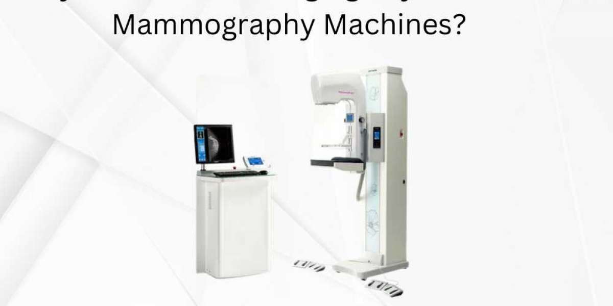
NOTE: Mammography Machine UAE was supplied and supported by Paramount Medical Equipment Trading LLC, ensuring clinics had access to advanced low-dose imaging technology and expert support. For reliable mammography solutions in the UAE, Paramount Medical Equipment Trading LLC has been the trusted partner for healthcare providers.
Why Radiation Dose Matters in Mammography
Understanding Radiation Risks
While mammograms use X-rays—a form of ionizing radiation—to create images of breast tissue, excessive exposure can increase the risk of developing cancer over time. Although the dose used in mammography is relatively low, minimizing exposure is crucial, especially for women who undergo regular screenings over many years. Low-dose imaging addresses this concern by reducing the cumulative radiation a patient receives, thereby lowering potential risks.
Balancing Safety and Diagnostic Accuracy
The challenge in mammography is to achieve the best possible image quality with the least amount of radiation. Too little radiation can result in poor image quality and missed diagnoses, while too much increases health risks. Modern low-dose mammography machines strike this balance by using advanced technology to optimize both safety and accuracy.
The Science Behind Low-Dose Mammography
Digital Detectors and Image Processing
Digital mammography systems utilize highly sensitive detectors that require less radiation to produce clear images. These detectors capture X-ray photons more efficiently than traditional film, allowing for lower doses without sacrificing detail. Advanced image processing algorithms further enhance image clarity, making it easier for radiologists to detect small tumors or subtle abnormalities.
3D Tomosynthesis and Composite Imaging
Three-dimensional mammography, or tomosynthesis, takes multiple low-dose images from different angles to create a detailed, layered view of the breast. This technique improves cancer detection rates, especially in women with dense breast tissue, and reduces the likelihood of false positives. Newer technologies, such as composite view (C-View), combine 3D and 2D images in a single low-dose exposure, reducing radiation by up to 40% and shortening the time required for breast compression.
Clinical Benefits of Low-Dose Imaging
Improved Cancer Detection
Low-dose 3D mammography has been shown to increase the accuracy of breast cancer screening. Studies reveal that tomosynthesis detects more invasive cancers and reduces the number of patients called back for unnecessary additional imaging or biopsies. This not only improves patient outcomes but also decreases anxiety and the burden of follow-up procedures.
Better Detection in Dense Breasts
Women with dense breast tissue face a higher risk of breast cancer and greater challenges in detection. Low-dose 3D mammography provides clearer images and better contrast, making it easier to identify tumors that may be hidden in dense tissue. This technology is especially beneficial for early detection, when treatment is most effective.
Fewer False Positives and Callbacks
A major concern in breast cancer screening is the rate of false positives—cases where a mammogram suggests cancer, but further testing reveals none. Low-dose imaging, particularly with tomosynthesis, has been proven to lower false positive rates by around 15% compared to traditional digital mammography. This reduces unnecessary stress and invasive procedures for patients.
Enhanced Patient Comfort
Modern low-dose mammography machines are designed for efficiency and comfort. With faster imaging times and reduced compression duration, patients experience less discomfort during the procedure. The ability to capture both 2D and 3D images in a single low-dose exposure streamlines the process and improves the overall experience.
Technological Innovations Supporting Low-Dose Imaging
Advanced X-Ray Tubes and Detectors
The latest mammography machines use X-ray tubes that are finely tuned for breast imaging, delivering precise doses tailored to the patient’s breast size and density. Digital detectors are engineered for maximum sensitivity, ensuring high-quality images at the lowest possible dose.
Computer-Aided Detection (CAD)
CAD systems assist radiologists by highlighting areas of concern on mammograms. These systems use sophisticated algorithms to analyze images and flag potential abnormalities, increasing diagnostic accuracy while supporting the use of lower radiation doses.
Automated Exposure Control
Automated exposure control (AEC) technology adjusts the X-ray dose in real time based on the thickness and composition of the breast tissue. This ensures optimal image quality for each patient while minimizing unnecessary exposure.
Addressing Myths and Concerns About Low-Dose Mammography
Is Low-Dose Mammography Less Effective?
A common misconception is that reducing radiation dose might compromise the ability to detect cancer. In reality, advances in detector sensitivity and image processing have made it possible to achieve excellent diagnostic performance at lower doses. Clinical studies confirm that low-dose mammography is as effective—if not more so—than traditional methods in detecting breast cancer.
Does 3D Mammography Mean Higher Radiation?
While early versions of 3D mammography raised concerns about increased radiation, modern systems have overcome this challenge. Newer machines generate 3D and 2D images from the same low-dose data set, keeping total exposure comparable to or even lower than that of standard 2D mammography.
The Role of Low-Dose Imaging in Breast Cancer Prevention
Supporting Regular Screening
The primary goal of mammography is to detect breast cancer early, when it is most treatable. By minimizing radiation exposure, low-dose imaging encourages more women to participate in regular screenings without fear of cumulative risk. This leads to earlier detection, less aggressive treatments, and better survival rates.
Reducing Healthcare Costs
By lowering the number of false positives and unnecessary biopsies, low-dose mammography reduces the overall cost of breast cancer screening programs. Fewer callbacks and follow-up procedures mean less time, stress, and expense for both patients and healthcare systems.
The Future of Low-Dose Mammography
Integration with Artificial Intelligence
Artificial intelligence is poised to further enhance low-dose mammography by improving image analysis, reducing interpretation errors, and personalizing screening protocols. AI-driven tools can help radiologists identify subtle changes over time, supporting even earlier detection with minimal radiation exposure.
Expanding Access and Awareness
As low-dose imaging technology becomes more widely available, its benefits will reach broader populations, including those in underserved regions. Ongoing education and outreach are essential to ensure that all women understand the importance of regular, safe mammography screening.

Conclusion
Low-dose imaging is the key to modern mammography machines, offering a powerful combination of patient safety, diagnostic accuracy, and comfort. By reducing radiation exposure without compromising image quality, these technologies are setting new standards in breast cancer screening and prevention. As innovation continues, low-dose mammography will remain at the forefront of efforts to save lives and improve women’s health worldwide.
For More Insightful Articles Related To This Topic, Feel Free To Visit: wutdawut