If you're in a lab setting—whether academic, industrial, or clinical—getting accurate data from your protein analysis is non-negotiable. You’re likely measuring protein concentration for various applications, from drug development to food quality control. But are you ensuring precision? If not, you're risking inconsistent results, missed targets, and possibly failed experiments.
That’s where SDS PAGE (Sodium Dodecyl Sulfate–Polyacrylamide Gel Electrophoresis) becomes your go-to tool. Used effectively, it gives you clear, reproducible profiles of protein concentration and molecular weight, ensuring your data tells the truth every time.
Let’s break it down so you can apply SDS PAGE confidently to your next protein concentration assessment—and finally get the accurate profiling you’ve been aiming for.
Why You Need Accurate Protein Profiling
You already know that proteins play a central role in biological processes. But unless you quantify them precisely, you’re working with assumptions. Whether you’re studying protein expression in research or monitoring contaminants in pharmaceuticals, accurate profiling supports:
- Batch consistency
- Regulatory compliance
- Functional analysis
- Product development
- Scientific reproducibility
When you combine protein concentration estimation with SDS PAGE, you’re not just looking at numbers—you’re visually confirming what’s present and how much is there.
What Makes SDS PAGE Ideal for Protein Concentration
Unlike other protein quantification methods, SDS PAGE allows you to visualize individual proteins while estimating their relative concentrations.
When you prepare your samples properly and run them through a polyacrylamide gel, proteins separate based on molecular weight. The use of SDS ensures they carry a uniform negative charge, allowing size—not charge or shape—to be the only factor during migration.
Why this matters to you:
- You can assess purity and degradation.
- You can detect unexpected protein bands.
- You can compare concentrations across different samples visually.
If you're aiming for accurate profiling, SDS PAGE lets you see more than just a concentration number—it shows you what that number really means in molecular terms.
Sample Preparation: The Foundation of Accuracy
Getting your sample prep right is the first step to success. Here’s a checklist you should follow before running SDS PAGE:
- Use a reliable lysis buffer to extract proteins without degradation.
- Quantify your protein concentration using a spectrophotometric assay like BCA or Bradford before gel loading.
- Normalize your samples to ensure each lane is loaded with the same total protein concentration.
- Add SDS and reducing agents (like DTT or β-mercaptoethanol) to denature the proteins completely.
This step sets the tone for how well your SDS PAGE will perform. A poorly prepared sample won’t just give bad results—it’ll mislead you entirely.
For detailed sample prep protocols tailored to various applications, you can look at this web-site which compiles reliable methods from experienced researchers.
Running the SDS PAGE Gel: What You Should Watch For
Once you’ve prepared your samples, it’s time to run the gel. Here’s how to ensure you're on the path to accurate profiling:
- Choose the correct gel percentage: Use lower percentages (e.g., 8%) for large proteins and higher (e.g., 15%) for small proteins.
- Load consistent volumes: Always use equal amounts of protein and loading buffer to maintain comparability.
- Run the gel at a stable voltage: Avoid high voltages that can cause smearing or uneven migration.
You'll want to include a protein ladder in one lane. This acts as a molecular ruler, helping you identify unknown protein sizes and cross-check against your expected band positions.
Staining and Imaging: Making Proteins Visible
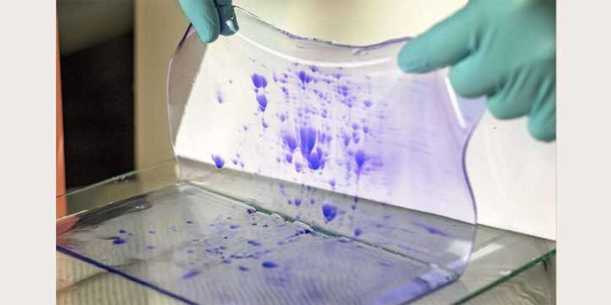
Once your gel is run, staining is your next step. Coomassie Brilliant Blue is a standard, but silver staining and fluorescent dyes offer greater sensitivity if you're working with low protein concentrations.
Here’s what you gain from proper staining:
- Visual confirmation of protein quantity via band intensity
- Identification of protein degradation or contamination
- Image archiving for documentation or publication
Use imaging software to quantify band intensity. The darker the band, the more concentrated the protein. Comparing the intensity of your sample bands to known standards allows you to estimate protein concentration semi-quantitatively, with visual and digital support.
Analyzing Your Results with Confidence
With your gel imaged, now you interpret the results. You should:
- Measure band density using densitometry tools.
- Compare with standard curves prepared from known protein concentrations.
- Normalize results to account for loading differences if needed.
If done correctly, your SDS PAGE gel becomes more than just a visual—it becomes a quantitative tool to track your proteins in a variety of biological samples.
If you're looking to automate this process or apply high-throughput techniques, learn more here about available software and image analysis platforms designed specifically for SDS PAGE quantification.
Common Pitfalls and How to Avoid Them
Even if you're experienced, SDS PAGE can go wrong. Watch out for:
- Smearing bands – usually from overloaded samples or degraded proteins
- Faint bands – possibly due to low protein concentration or poor staining
- Uneven migration – from buffer inconsistencies or gel defects
To avoid these, always:
- Run controls
- Use fresh reagents
- Monitor voltage and temperature during electrophoresis
- Calibrate pipettes regularly
Avoiding these common issues keeps your protein concentration profiling accurate and trustworthy.
When to Use SDS PAGE Over Other Methods
You might be asking yourself—why not just stick to colorimetric assays for concentration? The answer lies in the visual component.
SDS PAGE not only shows how much protein is there but what kinds of proteins are present. This is essential in:
- Recombinant protein expression
- Protein purification
- Allergen detection
- Biopharmaceutical product testing
In short, if your project requires both quantification and qualification of proteins, SDS PAGE should be your default.
Real-World Applications That Rely on You Getting It Right
You may be working on:
- Developing a new therapeutic protein
- Assessing the nutritional value of a dairy product
- Monitoring impurities in a vaccine
- Studying disease biomarkers
In all these cases, your ability to accurately measure protein concentration and profile it through SDS PAGE determines the reliability of your findings and the quality of your decisions.
So the next time you're setting up a gel, remember—you’re not just running a test. You’re ensuring accuracy in something that may affect health, safety, or innovation.
Final Thoughts
Protein concentration SDS PAGE isn’t just another lab technique. It’s a foundational tool in your toolbox—offering a visual and semi-quantitative look at the proteins that matter most in your research or product.
By preparing your samples carefully, running your gels consistently, staining and imaging properly, and analyzing the data precisely, you're giving yourself the edge in protein science.
If you're aiming for confidence and clarity in your results, SDS PAGE for protein concentration profiling is how you get there.
Original Source: https://kendricklabs.blogspot.com/2025/05/protein-concentration-sds-page-for.html


